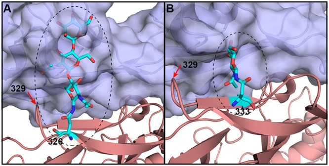
FIG 7.

Probable conformations of glycans linked to the indicated residues. bCD26s exhibit multiple glycosylation patterns at the receptor-ligand interface. Based on the structure of M. davidii bCD26 in complex with MERS-RBD, the possible positions and conformations of residue 326- and 333-linked glycans (numbering in M. davidii bCD26) are displayed. (A) Possible glycans on residue 326; (B) possible glycans on residue 333. Certain bCD26s (e.g., P. alecto ) and P. vampyrus) contain glycans linked to residue 329, which is highlighted with the red arrow in A and B.
