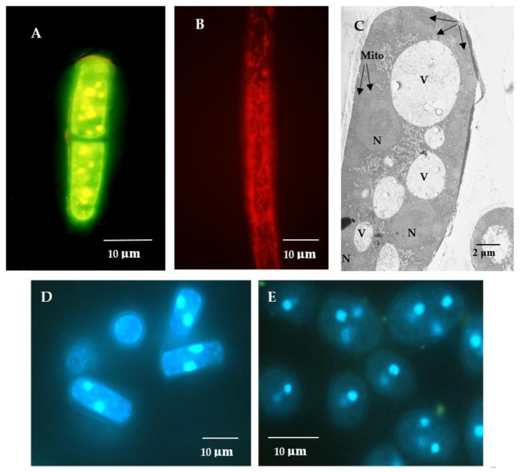
Figure 1.
Potential-dependent staining of mitochondria in the E. magnusii cells raised in the logarithmic growth phase by JC1 (A), Rh123 (B). (D,E)—the cells were raised in the logarithmic (D) and late stationary stages (E) and labeled with 0.3 μM DAPI for DNA. A,B—the cells were incubated with 0.5 μM JC1 or Rh123 for 20 min. The incubation medium contained 0.01 M phosphate-buffered saline (PBS), 1% glycerol, pH 7.4. The areas of high mitochondrial polarization are indicated by bright-yellow (A), bright-red (B) fluorescence due to the concentrated dye. To examine the Rh123-stained preparations, filters 02, 15 (Zeiss) were used (magnification 100×). Photos were taken using an AxioCam MRc camera. C—ultrastructure of the E. magnusii cells after 30 min incubation. N—nucleus, Mito—mitochondria, V—vacuole. (C)—ultrastructure of the E.magnusii cells. N—nuclei, V—vacuoles, Mito—mitochondria.

