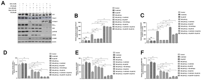
Figure 5.
Inhibition of delphinidin-induced autophagy increases H2O2-induced apoptosis in C28/I2 chondrocyte cells. (A) The C28/I2 chondrocyte cells were treated with 500 µM H2O2 in the presence or absence of 40 µM delphinidin (DP), 100 nM rapamycin (Rapa), and 20 µM chloroquine (CQ) for 4 h. After cell lysis, total cell extracts (30 µg) were separated on 8% or 10% SDS-PAGE and analyzed by Western blotting using primary antibodies against proteins (LC3, Bcl-XL, caspase-3, cleaved caspase-3, Nrf2, NF-κB, p-NF-κB, and cleaved PARP). β-Actin was used as a loading control. (B–F) Quantifications of protein expression and activation. The relative amounts of all proteins (Bcl-XL, caspase-3, cleaved caspase-3, Nrf2, NF-κB, p-NF-κB, and cleaved PARP, respectively) shown in Western blot analyses were quantified by NIH ImageJ software and represented as a graph. Data represent the means (± SD) of three independent experiments (* p < 0.05, ** p < 0.01; ns indicates not significant).

