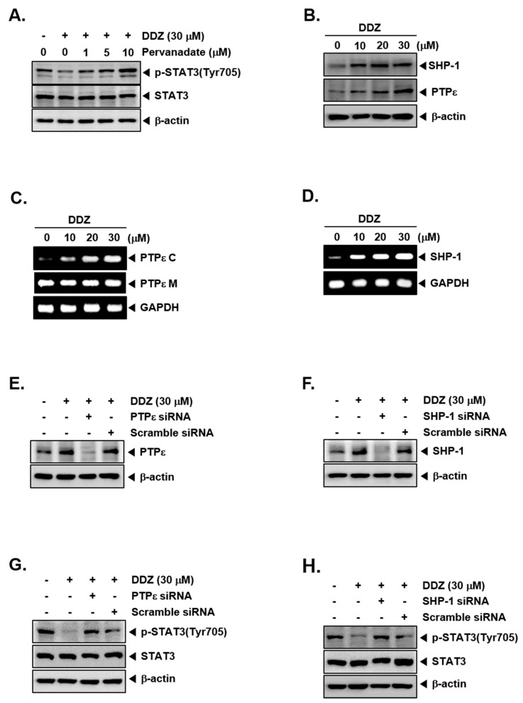
Figure 3.
DDZ increased protein tyrosine phosphatases protein tyrosine phosphatase (PTPε) and SHP-1 levels. (A) U266 cells (1 × 10⁶ cells/well) were treated with various concentrations of pervanadate and 30 μM of DDZ for 3 h. The expression of various proteins was analyzed by western blotting. (B) U266 cells (1 × 10⁶ cells/well) were treated with the indicated concentrations of DDZ for 3 h, and western blotting was done. (C) and (D) U266 cells (1 × 10⁶ cells/well) were treated as described above in panel (B), and total RNA was extracted and examined for the expression of various genes. (E)–(H) U266 cells (2 × 10⁶ cells/well) were transfected with scrambled or PTPε- or SHP-1-specific siRNA (100 nM). After 24 h, cells were treated with 30 μM of DDZ for 3 h, and the western blot analysis was done. The results shown are representative of three independent experiments.

