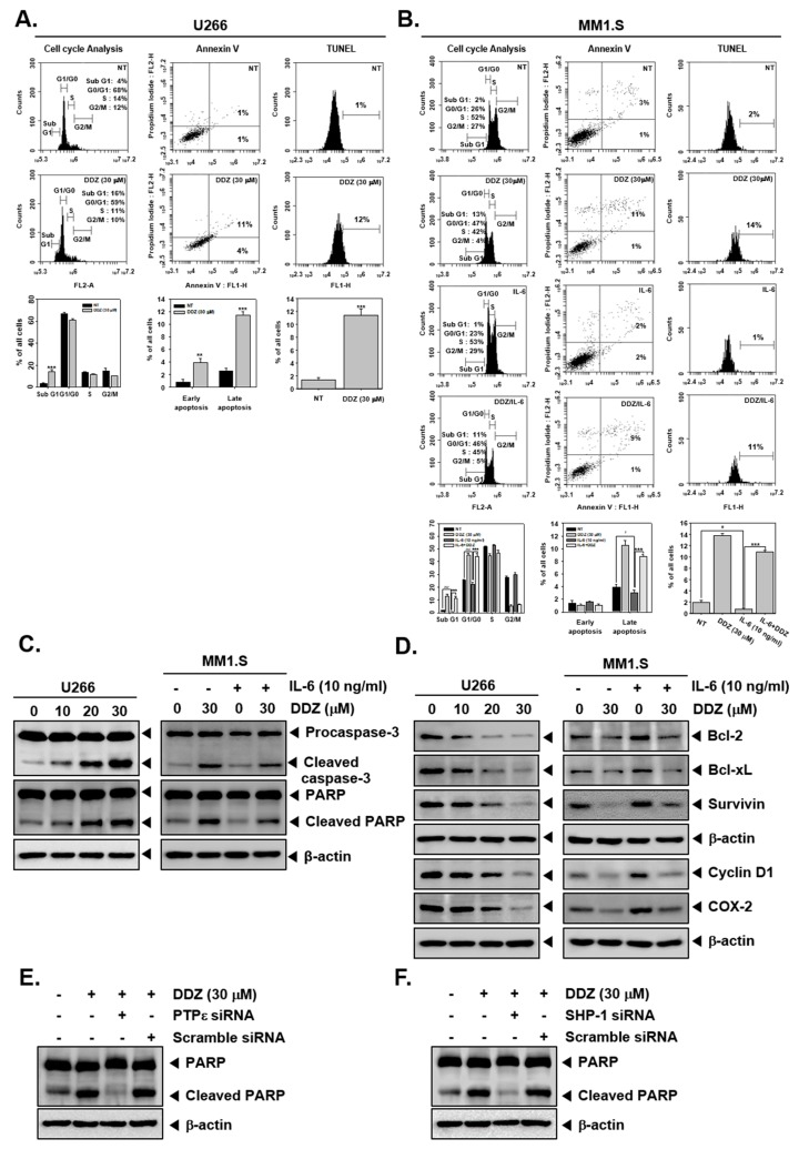
Figure 4.
DDZ promotes apoptosis in multiple myeloma (MM) cells. (A, B) U266 cells were treated with 30 μM of DDZ for 24 h, and MM1.S cells were treated with 30 μM of DDZ for 24 h and stimulated with IL-6 (10 ng/mL) for 10 min. Cell cycle analysis: The cells were fixed with 100% ethanol, and cell cycle analysis was done using cytometry. Annexin V: cells were stained with Annexin V FITC and PI for 15 min before being analyzed by flow cytometry. TUNEL: The cells were fixed by 4% PFA (paraformaldehyde), stained with a TUNEL assay reagent, and analyzed with flow cytometry. The results are presented as the mean ± SD. ** p < 0.01, *** p < 0.001, # p < 0.05, ### p < 0.001 compared the control. (C, D) U266 cells (1 × 10⁶ cells/well) were treated with indicated concentrations of DDZ for 24 h, and various proteins were examined by western blot analysis. MM1.S cells (1 × 10⁶ cells/well) were treated with 30 μM of DDZ for 24 h and stimulated with IL-6 (10 ng/mL) for 10 min. Then various proteins were examined by western blot analysis. (E, F) U266 cells (2 × 10⁶ cells/well) were transfected with scrambled or PTPε- or SHP-1-specific siRNA (100 nM). After 24 h, cells were treated with 30 μM of DDZ for 24 h. Whole cell lysates were prepared and analyzed by western blotting against the PARP protein.

