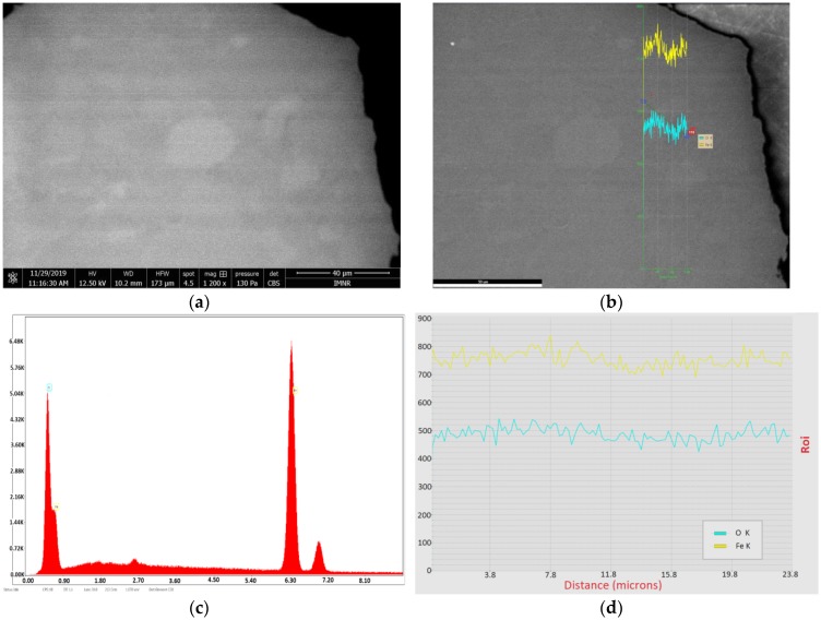
Figure 4.
Scanning Electron Microscopy with Energy Dispersive Spectroscopy (SEM-EDS) characterization of a hematite sample (NV4) prepared by hydrothermal synthesis at 20 bar, embedded in EpoThin-Buehler resin: (a) micrograph by scanning electron microscopy (SEM)collected using Circular Backscatter Detector (CBS); magnification 1200×; scale bar 40 µm; (b) line between hematite and goethite areas; (c) elemental analysis by energy dispersive X-ray spectroscopy (EDS); (d) distribution of O (turquoise) and Fe (yellow) content along the line between hematite and goethite areas from left to right.

