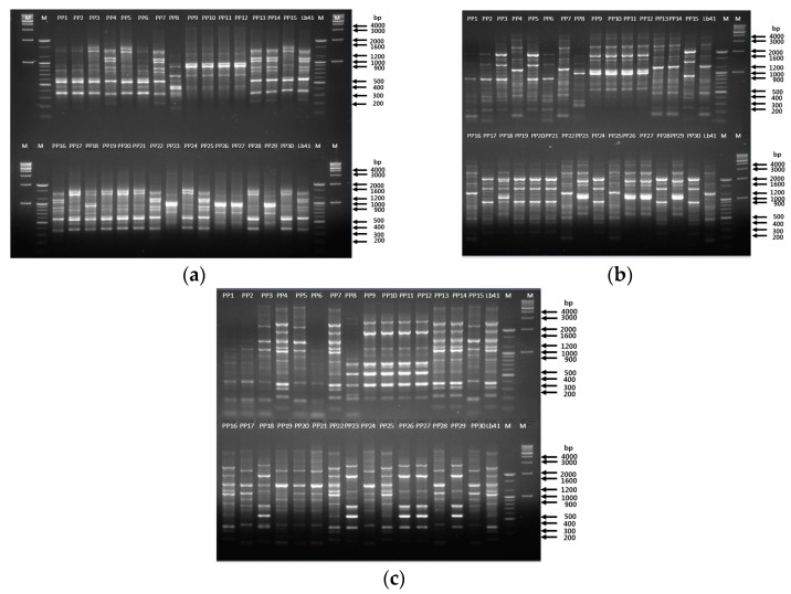
Figure 5.
Tracing of the suspected colonies from probiotic powder using rep-PCR analysis. (a) Primer (GTG)5; lane 1—marker 1 kb, lane 2—marker 100 bp, lane 3–17 (colonies PP1–PP15), lane 18—LB41P strain, lane 19—marker 100 bp, lane 20—marker 1 kb; lower half of the gel: lane 21—marker 1 kb, lane 22—marker 100 bp, lane 23–37 (colonies PP16–PP30), lane 38—LB41P strain, lane 39—marker 100 bp, lane 40—marker 1 kb. REP primer (b); ERIC primer (c); lane 1–15 (colonies PP1–PP15), lane 16—LB41P strain, lane 17—marker 100 bp, lane 18—marker 1 kb; lower half of the gel: lane 19–33 (colonies PP16–PP30), lane 34—LB41P strain, lane 35—marker 100 bp, lane 36—marker 1 kb. Positive control—LB41P.

