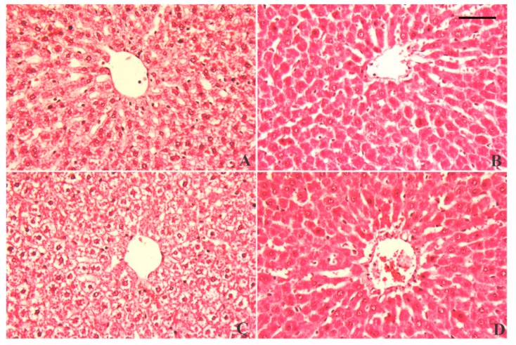
Figure 9.
Histopathological alterations of liver tissue following lead acetate (PbAc, 20 mg/kg, i.p.) and/or luteolin (LUT, 50 mg/kg, orally) exposure in male rats. Normal hepatic histological architecture was observed in the control and LUT groups, characterized by normal central veins surrounded by normal, intact hepatocytes (A and B, respectively). In contrast, PbAc-intoxicated rats exhibited necrotic liver cells associated with considerably degenerated and vacuolated peripheral hepatocytes, along with neutrophil and lymphocyte infiltrations around the peri-portal areas (C). However, pretreatment with LUT reversed the histological changes in response to PbAc intoxication (D). Scale bar 80 um.

