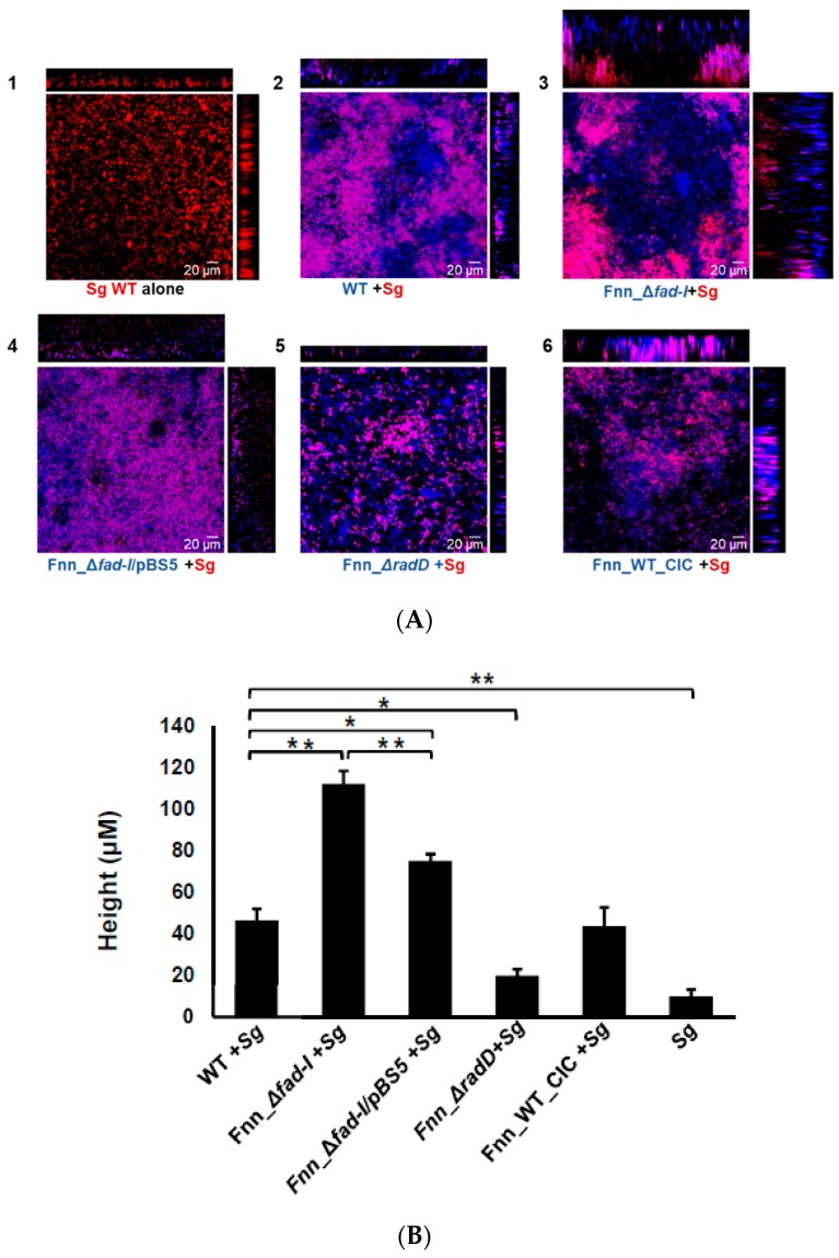
Figure 8.
(A). Visualization of dual-species biofilm formed between wild-type and mutant strains of F. nucleatum ssp nucleatum ATCC 23726 and S. gordonii (mCherry) by CLSM. The biofilm was fluorescently labeled with SYTO9. The S. gordonii (Sg) cells constitutively express mCherry and appear red on the images. Wild-type (WT) F. nucleatum (Fn) and its mutants (fad-I, fad-I/pBS5, radD, WT-CIC) are stained by syto9-only which are pseudo-colored as blue in the Zen software. Association of F. nucleatum and S. gordonii in the biofilm is observed as purple color in the confocal images. Each image panel is represented by x-z axis view on top and y-z axis view on the right side of the x-y view. The various panels show the biofilm formed by: (1) S. gordonii (Sg) alone; (2) wild-type ATCC 23726 with S. gordonii; (3) Fnn_Δfad-I with S. gordonii; (4) Fnn_Δfad-I/pBS5 with S. gordonii; (5) Fnn_ΔradD with S. gordonii; (6) Fnn_WT_CIC with S. gordonii. (B) Comparison of the height of the dual species biofilm of the wild-type and mutant strains of F. nucleatum ssp nucleatum ATCC 23726 with S. gordonii, as observed from the confocal images. The data represents mean of the height and standard error of mean of biofilm as observed in three independent experiments with height measurements captured in five randomly chosen locations in each experiment (n = 15). The single species biofilm of S. gordonii is also included as control. (* represents p ≤ 0.05, ** represents p ≤ 0.01).

