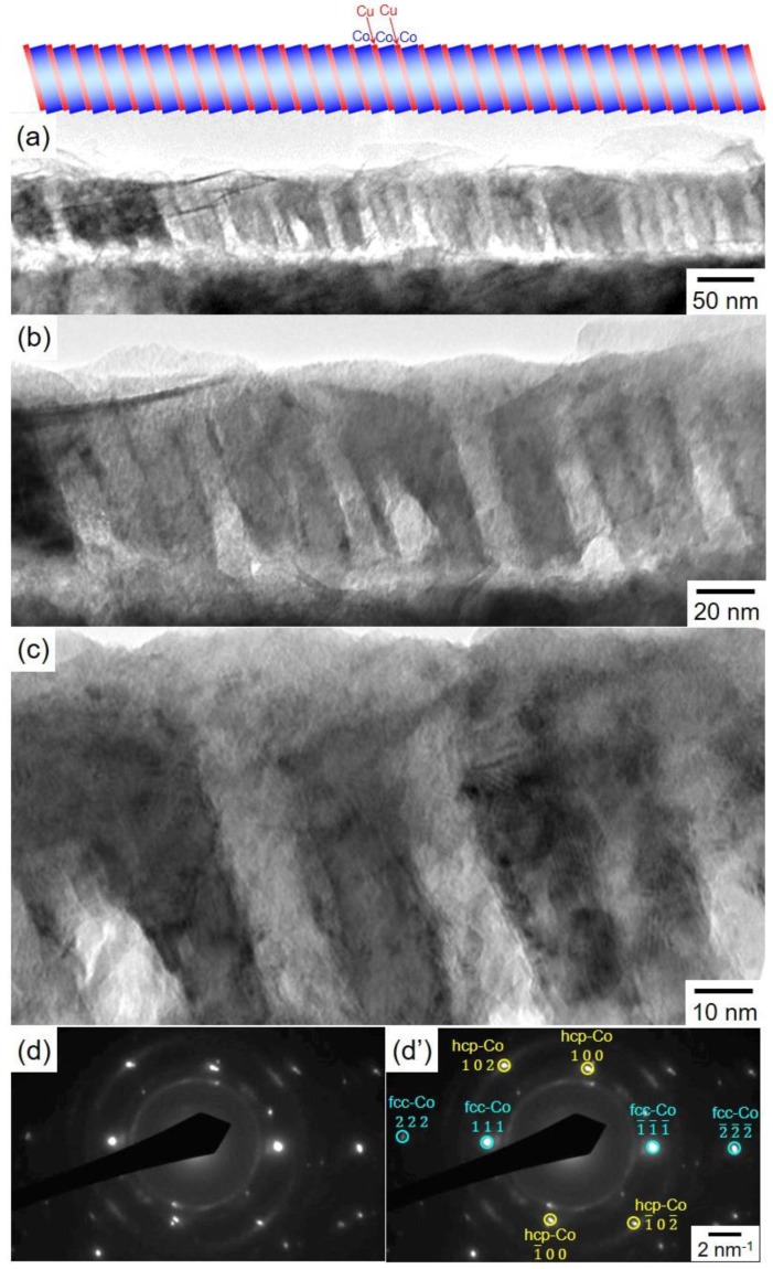
Figure 6.
TEM images of a Co/Cu multilayered nanowire that was pulsed-potential deposited at −0.40 V for 1.0 s and −1.05 V for 0.1 s (a–c). The electron diffraction patterns are also shown in (d–d’). The Co/Cu multilayered nanowires were separated from an anodized aluminum oxide nanochannel template.

