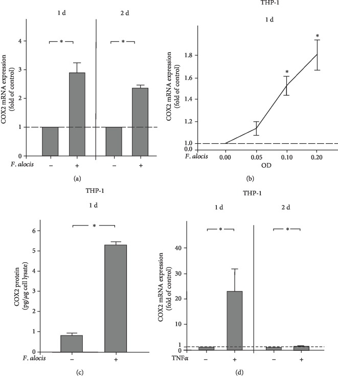
Figure 3.
(a) Expression of COX2 in the presence and absence of F. alocis (OD660: 0.1) in THP-1 cells at 1 d and 2 d, as analyzed by qPCR. Mean ± SEM (n = 18). ∗Significant (p < 0.05) difference between groups. (b) Stimulation of COX2 expression by various concentrations of F. alocis (OD660: 0.05, 0.1, and 0.2) in THP-1 cells at 1 d, as analyzed by qPCR. Unstimulated cells served as the control. Mean ± SEM (n = 18). ∗Significantly (p < 0.05) different from the control. (c) COX2 protein level in lysates of THP-1 cells in the presence and absence of F. alocis (OD660: 0.1) at 1 d, as analyzed by ELISA. Mean ± SEM (n = 18). ∗Significant (p < 0.05) difference between groups. (d) Expression of COX2 in the presence and absence of TNFα (1 ng/ml) in THP-1 cells at 1 d and 2 d, as analyzed by qPCR. Mean ± SEM (n = 12). ∗Significant (p < 0.05) difference between groups.

