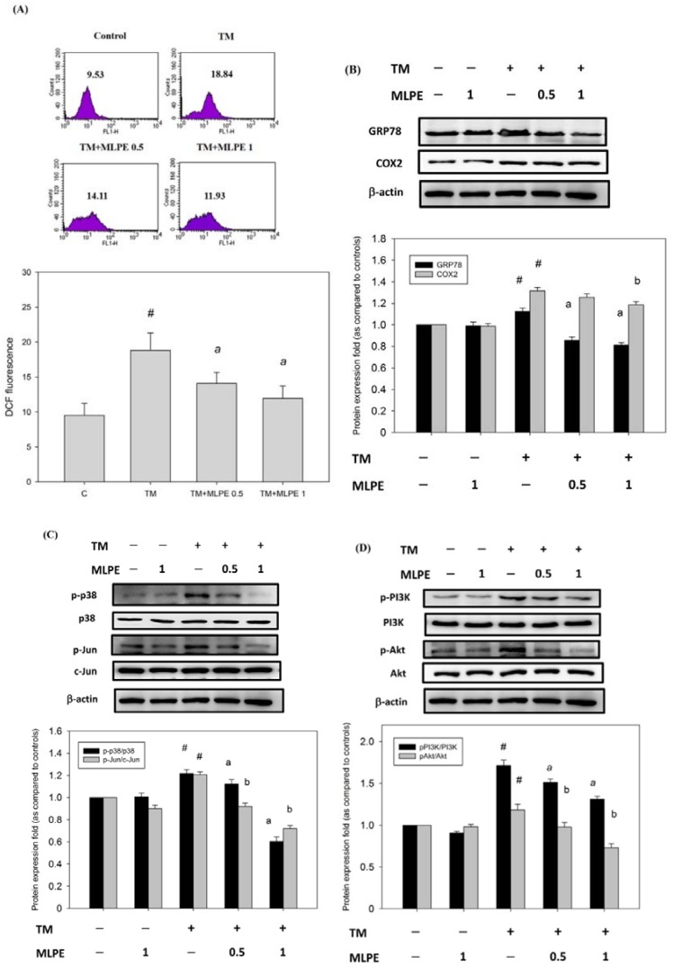
Figure 4.
MLPE reduced the level of reactive oxygen species (ROS) and the expression of COX-2, GRP78, p38/Jun, and PI3K/Akt in tunicamycin-induced HepG2 cells. HepG2 cells were treated with TM in the absence (control) or the indicated concentration of MLPE (0.5 and 1 mg/mL) for 8 h. (A) Intracellular ROS was detected by dichloro-dihydro-fluorescein diacetate (DCFH-DA) fluorescence. Data were analyzed with flow cytometry. Quantitative assessment was carried out of the DCF fluorescence cell. (B) COX-2 and GRP78; (C) p38/Jun pathway; (D) PI3K/Akt pathway were assessed through Western blotting. Whole cell lysates were subjected to Western blotting analysis. β-actin in the same HepG2 cell extract was used as an internal reference. Optical density reading values of the specific protein versus the loading control protein β-actin are represented as fold of the control values. # p < 0.05, compared with untreated HepG2 cells. a, b, p < 0.05, compared with HepG2 pretreated with TM for 8 h. +, means cell treated with the indicated chemical; −, means cell treated without the indicated chemical.

