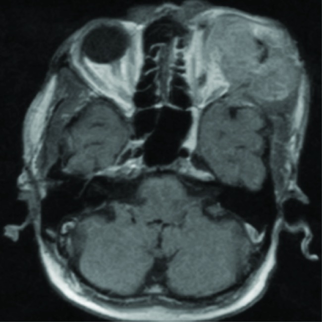
Fig.2.

Computed tomography of head and neck revealing heterogeneously enhancing irregular extraconal mass lesion measuring approximately 5.7 × 4.7cm in the superolateral aspect of the left orbit. It is breaching the lateral wall and roof of the orbit and extending into subcutaneous scalp tissue layers of the left frontal region. It is causing mass effect on eyeball resulting in the proptosis of the left eyeball and invading left-sided extraconal muscles, left lateral rectus, and superior oblique muscles.
