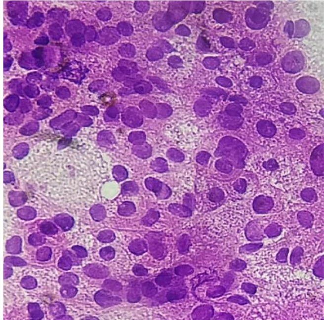
Fig.4.

Fine needle aspiration cytology smears from proptotic lesion and liver massshow individually scattered, clusters, and sheets of round to polyhedral cells with enlarged, hyperchromatic nuclei, prominent nucleoli, and moderately abundant eosinophilic cytoplasm. Anisonucleosis is also seen. Some cells show intranuclear inclusions. Many atypical stripped nuclei are seen. Scattered cells with large, bizarre nuclei are seen.
