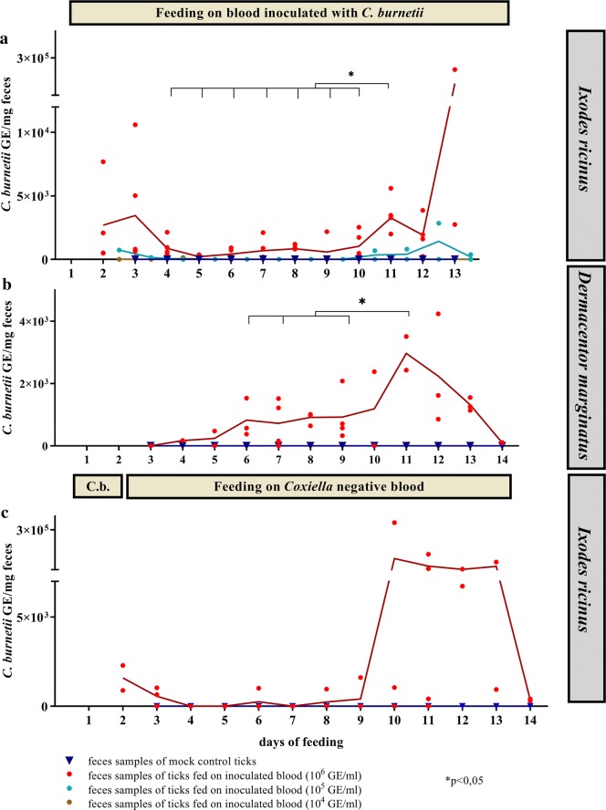
Fig. 2.
Coxiella DNA in feces. In (a) and (b) ticks were fed during the whole feeding experiment with blood inoculated with C. burnetii in different concentrations. The means of the replicates of each data set are connected with a line. The data for I. ricinus (a) represent three independent experiments with five different feeding units inoculated with 106 GE/ml blood (n = 5), 104 GE/ml (n = 2), 105 GE/ml (n = 2) and negative controls (n = 2). For D. marginatus (b), the data represent two independent experiments with feeding units on blood inoculated with 106 GE/ml (n = 4) and negative controls (n = 2). c Ticks were fed on 106 GE/ml inoculated blood for 36 h (indicated by the horizontal bar labelled with ‘C.b.’) (n = 4). Attached ticks were subsequently removed and placed in a Coxiella-free feeding unit. Ticks constantly feeding on Coxiella-negative blood served as negative controls (n = 2). Statistical significance of DNA loads in comparison to day 11 is indicated by an asterisk

