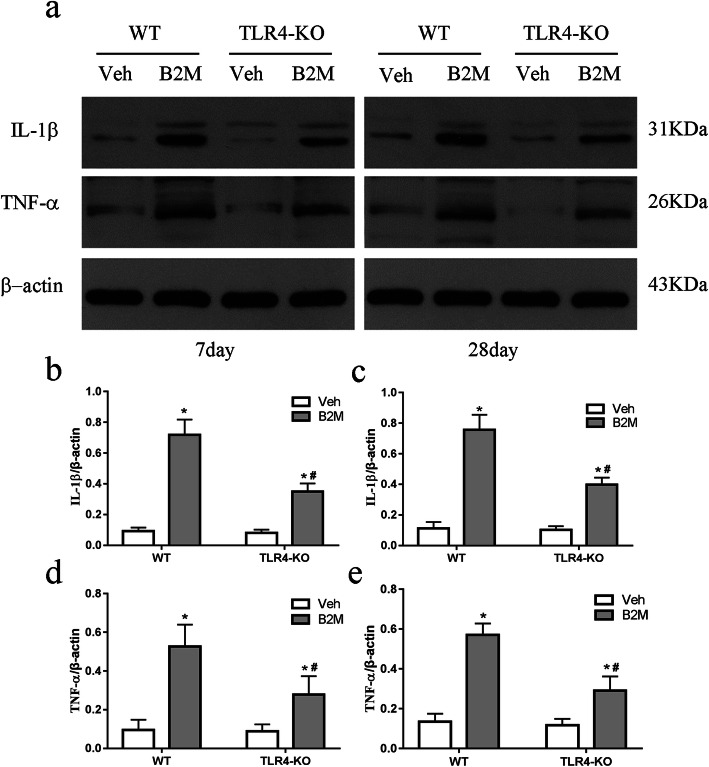
Fig. 5.
TLR4 elimination decreased the expression of IL-1β, TNF-α following B2M treatment. IL-1β and TNF-α protein expression in the hippocampus were determined at 7 and 28 days after B2M or vehicle treatment (n = 6/subgroup). The relative level in arbitrary units compared to β-actin (a). B2M treatment increased the levels of IL-1β and TNF-α were at 7 (b, d) and 28 days (c, e), which was reversed in TLR4-KO mice. The data were analyzed using two-way ANOVAs used Tukey’s multiple comparisons test. Values are presented as the means ± SD. Significant differences are expressed as follows: *p < 0.05 compared B2M group vs. Veh group in WT and TLR4-KO mice, according to the two-way ANOVA; #p < 0.05 compared B2M group in TLR4-KO mice vs. WT mice, according to the two-way ANOVA. IL-1β = interleukin-1β, TNF-α = tumor necrosis factor-alpha, WT = wild type C57BL/6, TLR4-KO = TLR4 knockout

