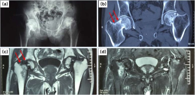
Fig. 2.
Imaging examinations of the patient in the hip region after admission into our hospital. a Plain radiograph of the pelvis indicating fracture of left femoral neck, with obvious displacement, nonunion and femoral neck shortening, and old fracture of the right femoral neck. b CT image of the coronal plane revealing bilateral femoral neck insufficiency fractures, with obvious displacement, nonunion and femoral neck shortening in the left femoral neck and double fracture line (arrows) in his right femoral neck. c Coronal T1-weighted image showing low signal intensity in the fracture region of bilateral femoral neck, with double fracture lines (arrows) on the right side. d Coronal T2-weighted image showing swelling on the right femoral neck and interruption of cortex on bilateral femoral neck

