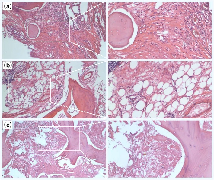
Fig. 4.
The HE staining pictures of bone tissue removed from the fracture region of the left femoral neck. a Some new but very few new bone tissues, with massive fibrous tissues, could be observed in the removed necrotic bone tissues. b some thinning trabeculae structure presented in some areas of the removed necrotic bone tissues. c Necrotic and fibrous bone tissues presented in most of the areas in the removed necrotic bone tissues. (magnification: 40 times)

