Fig. 3.
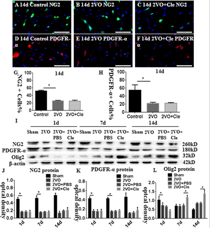
Immunofluorescence staining showing platelet-derived growth factor receptor (PDGFR)-α and neural/glial antigen 2 (NG2) immunoreactive oligodendrocytes (PDGFR-α, red; NG2, green) in the corpus callosum of rats at 14 days after the bilateral common carotid artery occlusion (BCCAO; B and E) and clemastine injection after BCCAO (c and f) when compared with their corresponding controls (a and d). Bar graph in G and H shows a remarkable decrease in the ratios of PDGFR-α and NG2-positive oligodendrocytes/mm2 in the corpus callosum at 14 days after the BCCAO when compared with their corresponding controls; clemastine showed no effect on the ratios of NG2 and PDGFR-α. Panel I shows NG2 (260 kDa), PDGFR-α (180 kDa), oligodendrocyte transcription factor 2 (Olig2; 32 kDa), and β-actin (42 kDa) immunoreactive bands. Panels J and K show bar graphs depicting significant decreases in the optical density of NG2 and PDGFR-α at 1, 7, and 14 days following the BCCAO when compared with their corresponding controls; clemastine showed no effect on the expression of NG2 and PDGFR-α. Panel L shows a bar graph depicting a significant decrease in the optical density of Olig2 at 1 day and increase at 7 and 14 days following the BCCAO when compared with their corresponding controls; Olig2 protein increases significantly at 7 and 14 days after treatment with clemastine compared with that of BCCAO. N = 3. *P < 0.05. Scale bars: A–F 50 μm.

