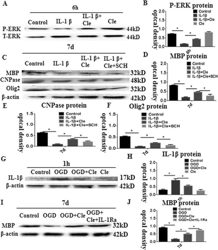
Fig. 6.
Western blot analysis showing clemastine-upregulated MBP, CNPase, and Olig2 expression via activation of extracellular regulated protein kinases (ERK) pathway in OPCs. Panel a showing ERK phosphorylation and total ERK immunoreactive bands. Panel b is a bar graph showing significant decreases in the optical density of ERK phosphorylation following treatment with IL-1β; clemastine reversed these changes. Panel c shows immunoreactive bands, which indicate that expression of MBP, CNPase, and Olig2 was inhibited in the OPCs after IL-1β administration for 7 days. The clemastine may attenuate the increment. SCH772984 which is an ERK pathway inhibitor could reverse the expression of MBP, CNPase, and Olig2 induced by IL-1β + clemastine treatment. Panels d–f are bar graphs showing remarkable suppression of MBP, CNPase, and Olig2 by SCH772984. Panel G shows immunoreactive bands, which indicate that expression of IL-1β was increased in the AMCs after oxyglucose deprivation (OGD) administration for 1 h; clemastine could remarkably reduce the expression of IL-1β (h). Panel i shows immunoreactive bands, which indicate that expression of MBP was decreased in the co-culture of microglia with OPCs at 7 days after oxyglucose deprivation (OGD) administration for 1 h; clemastine could upregulate the expression levels of MBP after OGD. When an IL-receptor antagonist was added, the expression of MBP increased compared with that in the clemastine group after OGD (J). N = 3. *P < 0.05

