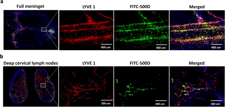
Fig. 2.
The subdural fluorescent macromolecule drainage pathway colocalized with lymphatics in the meninges and dCLNs. a Immunofluorescence images of mouse mLVs (LYVE 1+). The merged channel shows that FITC-500 D colocalizes with LYVE 1+ mLVs near the superior sagittal sinus. b Immunofluorescence images of the mouse lymph sinus of deep cervical lymph nodes (LYVE 1+). Fluorescence colours of (a) and (b): LYVE 1, red; FITC-500 D, green; DAPI, blue. n = 3 (mice). The merged channel shows that FITC-500 D appears in the lymph sinus

