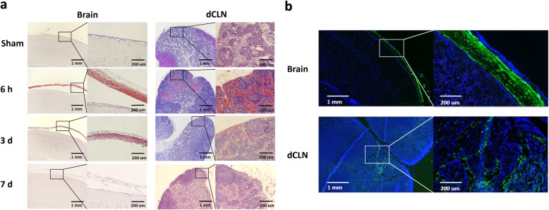
Fig. 3.
SDH drained into dCLNs. a H & E staining images of brain coronary and dCLN sections in the sham and SDH groups at 6 h, 3 d and 7 d after SDH establishment. n = 3/group. b Fluorescence images of rat brain coronary and dCLN sections at 6 h after the injection of A488-fibrinogen mixed with autologous blood into the subdural space. Green fluorescence signals were detected in the rat subdural space and dCLNs. Fluorescence colours: A488-fibrinogen, green; DAPI, blue. n = 3

