Figure 1.
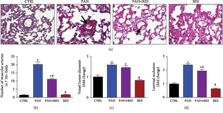
There is a limited effect of RES exerted in the lung vessel histopathology structure. (a) Representative microphotographs of pulmonary blood vessels. PAH induced hypertrophy and proliferation of the tunica media; this effect is decreased by RES. 20x magnification; H&E staining. Arrows indicate the muscularized vessel wall. (b) Amount of muscular arteries in 7 random fields in lung tissue. (c) Diameter of pulmonary blood vessels. (d) Luminal occlusion by the media layer in lung arteries. The values are given as the mean and fold change ± SEM; ∗p < 0.05 vs. control; #p < 0.05 vs. PAH; n = 15 for CTRL, PAH, and PAH+RES; n = 11 for RES.

