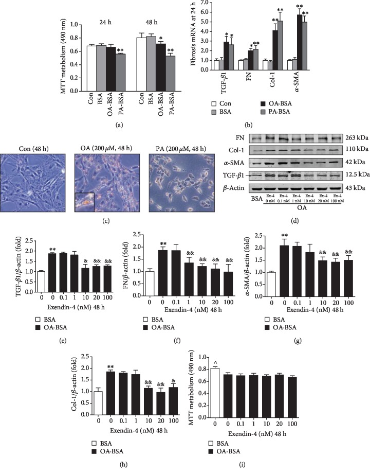
Figure 4.
Effect of FFA-BSA complexes on myofibroblast-like phenotype transition and lipid accumulation in cultured mesangial cells. Quiescent mesangial cells were treated with media (control, Con), 1% fatty acid-free BSA (BSA) alone, 200 μM oleate- or palmitate-1% fatty acid-free BSA (OA-BSA and PA-BSA, respectively), or exendin-4 of 0 (vehicle control), 0.1, 1, 10, 20, and 100 nM for 48 hours. (a) Proliferation of MCs by MTT assay. (b) Expressions of TGF-β1, FN, α-SMA, and collagen I by quantitative RT-PCR. (c) Lipid accumulation in MCs determined by Oil Red O assay. Inset presents ×10 magnification to illustrate size and location of lipid droplets in cytoplasm. (d) Western bolt analysis of TGF-β1, FN, α-SMA, and collagen I. Quantitative analyses of the results are also shown: TGF-β1 (e), FN (f), α-SMA (g), and Col-1 (h). Values are the mean ± SD of 3 independent experiments. ∗P < 0.05 and ∗∗P < 0.01 vs. BSA group, &P < 0.05 and &&P < 0.01 vs. OA-BSA with 0 nM exendin-4 group. (i) Dose response of MC viability in response to increasing concentration of exendin-4. ^P < 0.05 vs. other groups.

