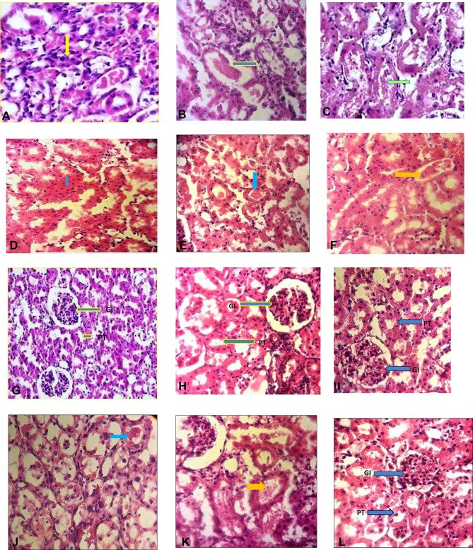
Figure 4.
The study of histopathologic analysis of the kidney of various tested rats after cisplatin induction. Histological section of cisplatin group; A (necrosis), B (cast hyaline), and C (intratubular debris). Histological sections of the kidneys of the control groups are normal and non-harmful (G). In the kidneys of the first experimental group, receiving DMEM + FBS medium (D, E, F), necrosis (green arrow), cast hyaline (blue arrow), and intra-tubular debris (yellow arrow) were found. In the kidneys of the second experimental group (H), receiving 5×10⁶ stem cells conditioned medium, it was found only Intra-tubular debris. In the kidneys of the third experimental group (I), receiving 5×10⁶ stem cells + GO, none of the damage was observed. In the kidneys of the fourth experimental group (J, K), receiving GO, there was found cast hyaline (J), and Intra-tubular debris (K). In the kidneys of receiving GO (without cisplatin injection), none of the damage was observed (L) and GO had no toxic effect. (H, E) staining × 400.

