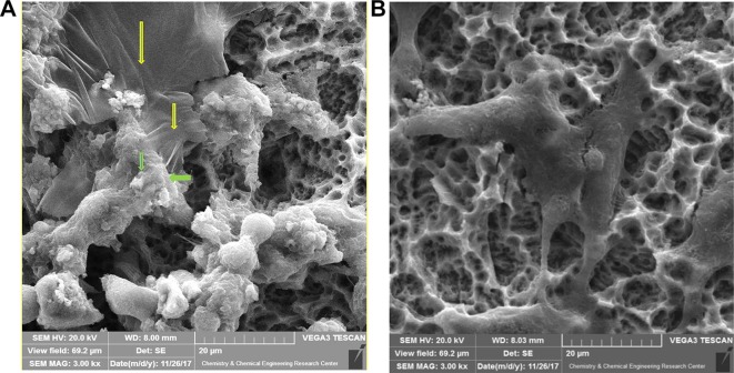
Figure 8.
SEM images of interactions among GO edges, MSCs, and growth factors derived from stem cells (A). Arrows indicate the attachment of stem cells and growth factors secreted from MSCs (green) to the edges of the silica-GO layer (yellow). In the absence of GO, the aggregation of MSCs was not sufficiently noticeable (B).

