
Figure 6. Cardiac fibroblasts from Thy1 KO mice express increased level of profibrotic genes in response to TAC.
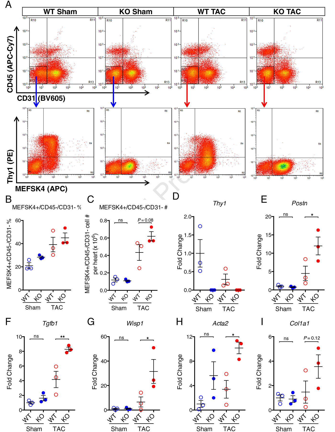
(A) Representative flow cytometry analysis of freshly isolated cells from WT and Thy1 KO mice in Sham or TAC conditions stained with APC-Cy7-conjugated CD45, BV605-conjugated CD31, PE-conjugated Thy1 and APC-conjugated MEFSK4. (B) Quantification of percentage fraction of MEFSK4+/CD45−/CD31− CFs in freshly isolated ventricular cells. (C) Quantification of MEFSK4+/CD45−/CD31− CFs cell number per heart (ventricles only) after FACS. Expression of Thy1 (D), Postn (E), Tgfb1 (F), Wisp1(G), Acta2 (H), and Col1a1 (I) genes in freshly isolated MEFSK4+/CD45−/CD31− CFs from WT and KO mice in Sham or TAC conditions. The mRNA expression was normalized to house-keeping gene B2M and shown as fold change relative to WT Sham group. N=3 independent experiments per group. ns: not significant, *P < 0.05, **P < 0.01 by 1-way ANOVA with Bonferroni’s multiple comparisons test.