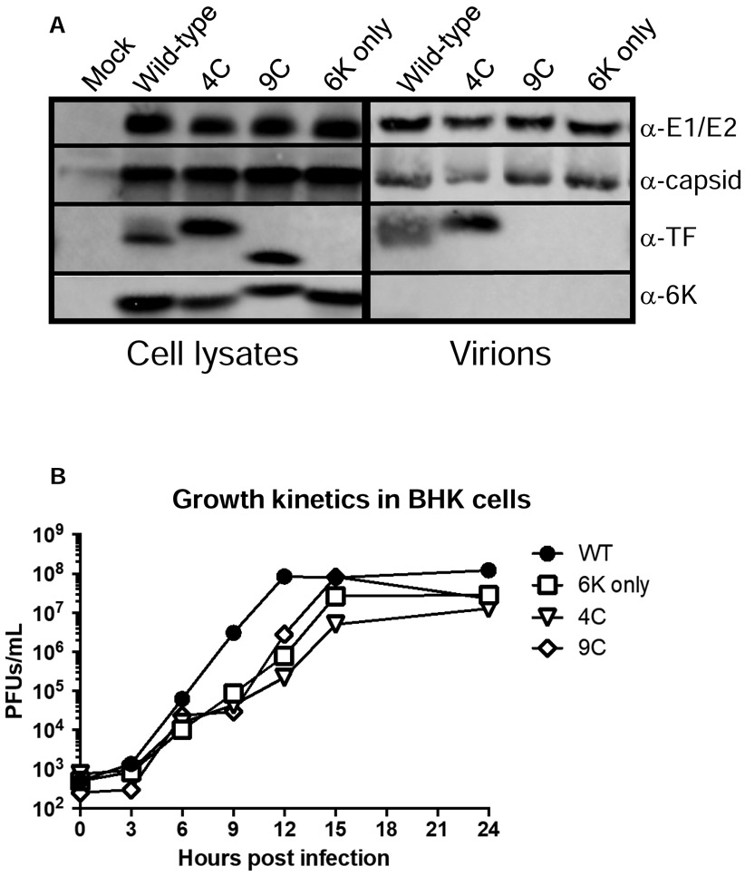
Figure 1: Characterization of viruses.
A) BHK cells were infected with wild-type or TF mutant of SINV (AR86) (MOI=1). At 16 hours post infection, cells were lysed and lysates probed with antibodies specific for E1/E2, capsid, TF and 6K proteins. To probe components in purified virions, BHK cells were infected (MOI=5) and after 24 hours, media was collected, clarified, and run through a sucrose cushion. Pellets were resuspended and virion associated proteins were separated by SDS PAGE followed by western blotting. B) Growth kinetics were evaluated in BHK cells by plaque forming assays. Confluent monolayers of BHK cells were infected with the indicated virus (MOI=5) for 1 hour at room temperature. Cells were washed to remove unbound virus, media was refreshed, and cells were shifted to 37°C. At the indicated timepoints, 200μl of supernatant were removed and titered on BHKs to determine viral concentrations. Titers are reported as plaque forming units per milliliter. Each point represents the mean of two independent experiments.

