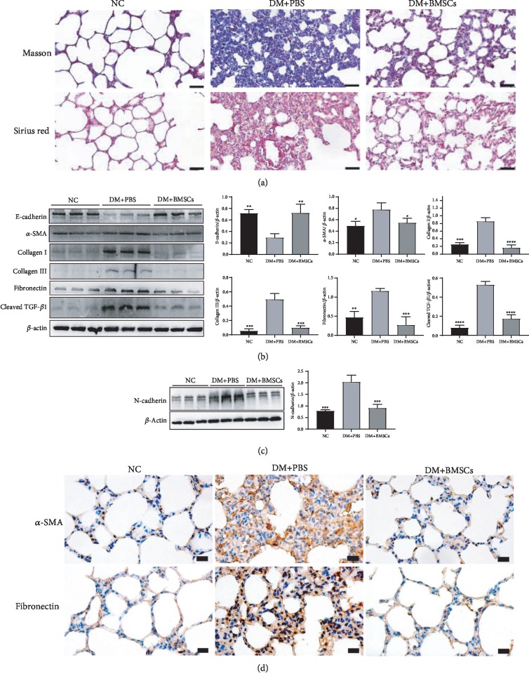
Figure 1.
MSCs inhibit lung fibrosis caused by diabetes in rats. (a) Masson and Sirius Red staining of lung tissues. Magnification, ×400. Scale bar, 50 μm. (b, c) Effects of MSCs on the protein expressions of E-cadherin, α-SMA, collagen I, collagen III, fibronectin, cleaved TGF-β1, and N-cadherin by the western blotting assay. (d) The representative micrographs of immunohistochemical staining of α-SMA and fibronectin. Magnification, ×200. Scale bar, 20 μm. Data are shown as mean ± standard deviation (∗P < 0.05, ∗∗P < 0.01, ∗∗∗P < 0.001, and ∗∗∗∗P < 0.0001 compared with the DM+PBS group, n = 6 per group).

