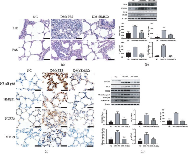
Figure 4.
MSCs attenuate lung inflammation in diabetic rats. (a) The representative micrographs of HE and PAS staining of the lung tissue. Magnification, ×400. Scale bar, 50 μm. (b) Effects of MSCs on the protein expressions of TNF-α, ICAM-1, IL-1β, and MMP9 in the lung tissue. (c) The representative micrographs of immunohistochemical staining of p65, HMGB1, NLRP3, and MMP9. Magnification, ×200. Scale bar, 50 μm. (d) Effects of MSCs on the protein expressions of HMGB1, RAGE, NLRP3, cleaved caspase-1, p65, and p-p65. Data are shown as mean ± standard deviation (∗P < 0.05, ∗∗P < 0.01, ∗∗∗P < 0.001, and ∗∗∗∗P < 0.0001 compared with the DM+PBS group, n = 6 per group).

