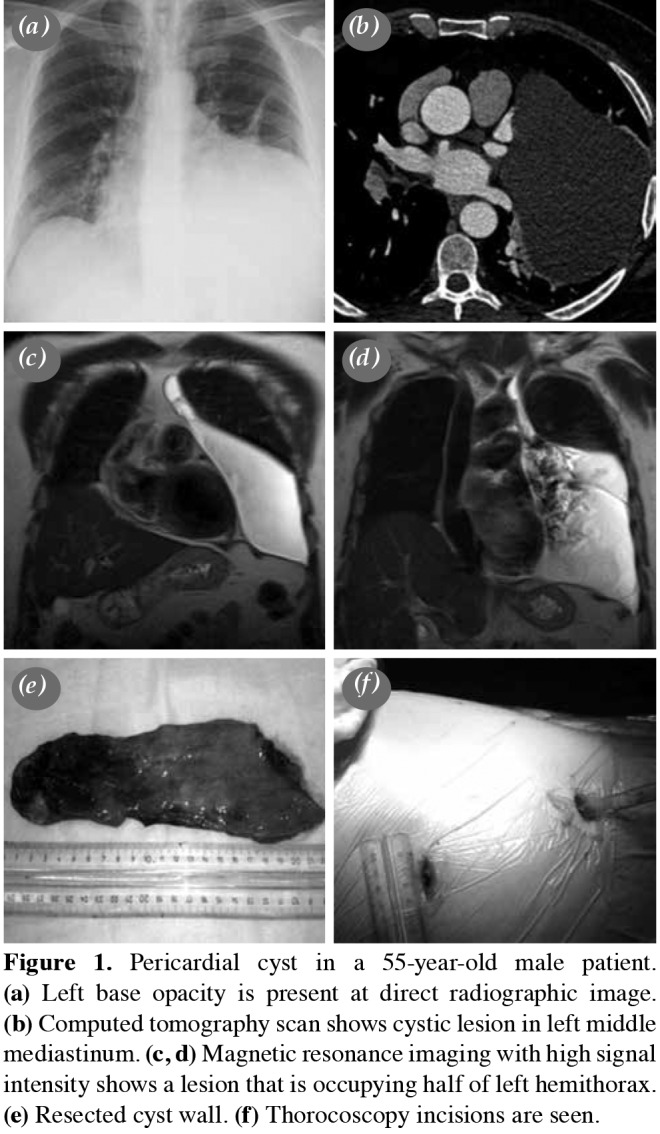
Figure 1. Pericardial cyst in a 55-year-old male patient. (a) Left base opacity is present at direct radiographic image. (b) Computed tomography scan shows cystic lesion in left middle mediastinum. (c, d) Magnetic resonance imaging with high signal intensity shows a lesion that is occupying half of left hemithorax. (e) Resected cyst wall. (f) Thorocoscopy incisions are seen.

