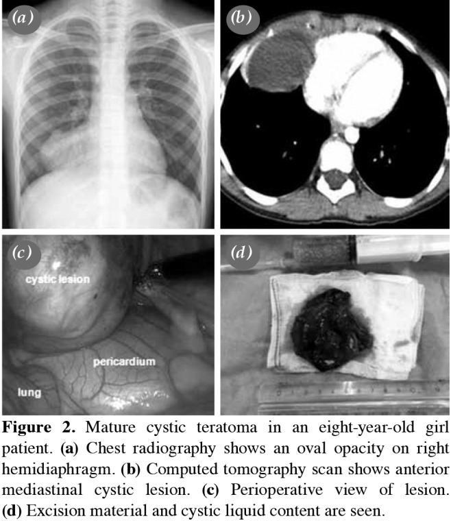
Figure 2. Mature cystic teratoma in an eight-year-old girl patient. (a) Chest radiography shows an oval opacity on right hemidiaphragm. (b) Computed tomography scan shows anterior mediastinal cystic lesion. (c) Perioperative view of lesion. (d) Excision material and cystic liquid content are seen.

