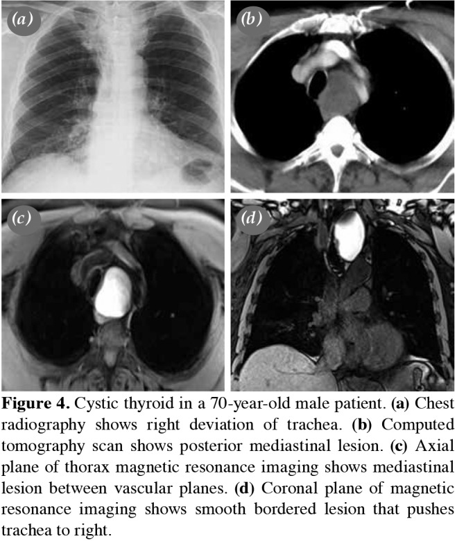
Figure 4. Cystic thyroid in a 70-year-old male patient. (a) Chest radiography shows right deviation of trachea. (b) Computed tomography scan shows posterior mediastinal lesion. (c) Axial plane of thorax magnetic resonance imaging shows mediastinal lesion between vascular planes. (d) Coronal plane of magnetic resonance imaging shows smooth bordered lesion that pushes trachea to right.