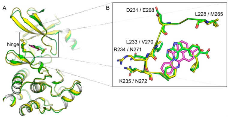
Figure 2.

(A) Superimposed structures of CAMKK1 (yellow, PDB 6CD6) and CAMKK2 (green, PDB 6BKU) bound to GSK650394. STO-609 (as bound to CAMKK2, PDB 2ZV2) is also shown in magenta. Spheres show positions where residues differ within the ATP-binding site of the two CAMKKs. (B) Inset shows a top view of the ATP-binding sites of both CAMKKs.
