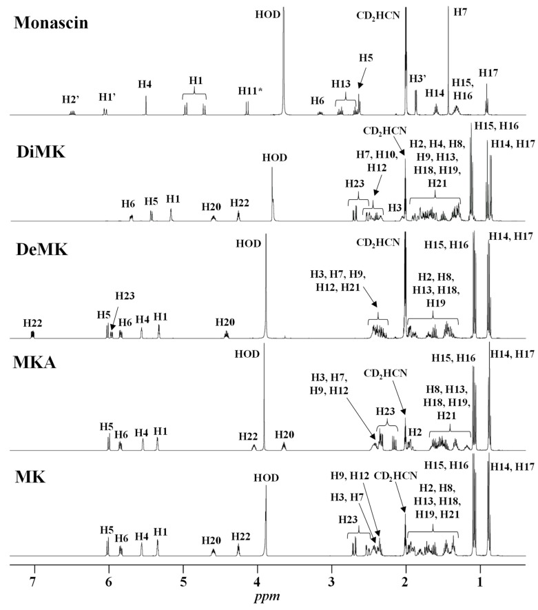
Figure 3.
1H-NMR spectra of standards of monacolin K in lactone form (MK) and in hydroxyl acid form (MKA), dehydromonacolin K (DeMK), dihydromonacolin K (DiMK) and monascin recorded in CD3CN:D2O (80:20). The chemical structures of all the compounds and their protons numbering are given in Table 2. (*) The signal of H11 of monascin disappears with time due to exchange with D2O.

