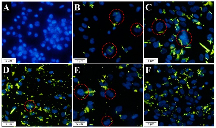
Figure 4.
Fluorescent microscopic images of DAPI-stained cells following 48-h treatment with A (fresh medium (control)), B (ligand L), C (complex Co2+), D (complex Ni2+), E (complex Cu2+), and F (complex Zn2+). Red circles indicate the Saos-2 nuclei with anomalous morphology after internalization of the fluorescent chemical compounds (L, and complexes of Co2+, Ni2+, Cu2+, and Zn2+).

