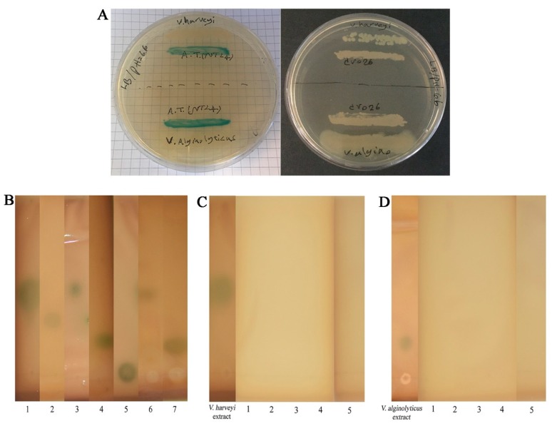
Figure 6.
Detection and degradation of natural AHLs. (A) Detection of AHLs produced by tested V. harveyi and V. alginolyticus by the cross-feeding assay. Induction of A. tumefaciens NTL4 (left plate) vs. non-induction of C. violaceum CV026 (right plate). (B) Thin-layer chromatography (TLC) analysis for tentative identification of AHLs extracted from tested V. harveyi (lane 6) and V. alginolyticus (lane 7). Synthetic AHLs were used as standards. Lane 1 (3-oh-C4-HSL), lane 2 (C6-HSL), lane 3 (3-oxo-C6-HSL), lane 4 (3-oxo-C10-HSL), and lane 5 (3-oxo-C14-HSL). (C–D) Visualization of the AHL-degrading activity of selected QQIs against natural AHLs of V. harveyi and V. alginolyticus using A. tumefaciens NTL4 overlay on TLC plates. The absence of blue color development in A. tumefaciens NTL4 indicates positive QQ activity. AHLs extracted from V. harveyi and V. alginolyticus cultures were regarded as negative controls. QQ1 (lane 1), QQ2 (lane 2), QQ3 (lane 3), QQ4 (lane 4), QQ5 (lane 5).

