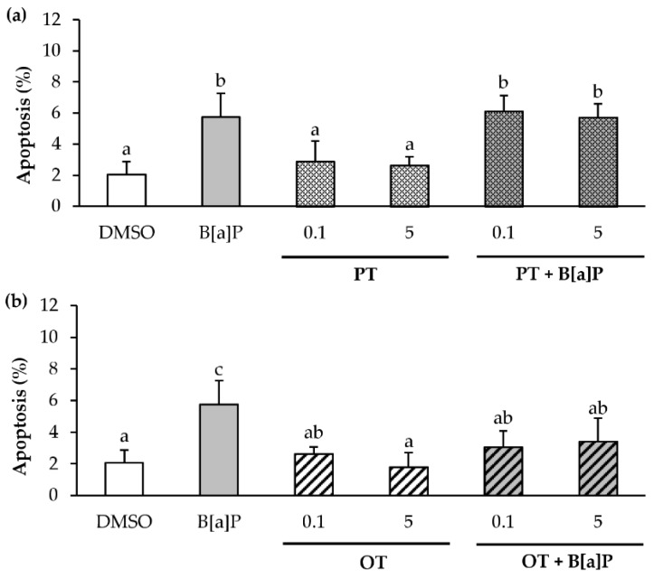
Figure 5.
Effects of P. tricornutum and O. tauri extracts on B[a]P-induced apoptosis in endothelial HMEC-1 cells. HMEC-1 cells were exposed to vehicle (DMSO) or treated with 2 μM B[a]P, or with 0.1, 1 and 5 µg/mL P. tricornutum (PT) extract (a) or O. tauri (OT) extract (b), or with a combination of the toxicant and the extract for 24 h. Apoptotic nuclei were analyzed by fluorescence microscopy after staining with Hoechst 33342. Results are represented as mean values ± SD from at least 3 independent experiments. Mean values assigned with different letters are significantly different (p < 0.05) with a < b < c.

