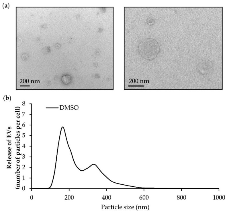
Figure 8.
Characterization of extracellular vesicles (EVs) released by endothelial HMEC-1 cells. HMEC-1 cells were exposed to DMSO for 24 h. EVs were isolated by differential ultracentrifugation. (a) Transmission electron microscopy pictures of EV pellets (scale bars = 200 nm). (b) Representative size distribution profile by nanoparticle tracking analysis (NTA) of EVs produced by endothelial cells.

