Figure 1.
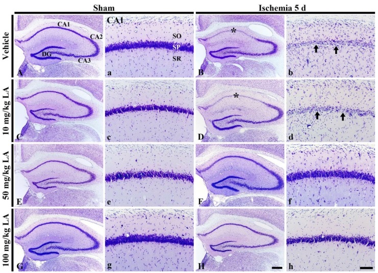
Cresyl Violet (CV) staining in the hippocampus (A–H) and its Cornu Ammonis 1 (CA1) field (a–h) of the vehicle/sham (A,a), 10, 50 and 100 mg/kg laminarin (LA)/sham (C,c,E,e,G,g), vehicle/ischemia (B,b) and 10, 50 and 100 mg/kg LA/ischemia (D,d,F,f,H,h) groups at 5 days after sham or transient forebrain ischemia (TFI) operation. In the vehicle/ischemia group, CV dyeability is remarkably reduced in the stratum pyramidale (SP, arrows) of the CA1 field (asterisks). In the 10 mg/kg LA/ischemia group, the distribution pattern of CV stained cells is similar to that in the vehicle/ischemia group. However, in the 50 mg/kg and 100 mg/kg LA/ischemia groups, CV stainability is conserved. DG, dentate gyrus; SO, stratum oriens; SR stratum radiatum. Scale bars = 400 μm (A–H) and 100 μm (a–h).

