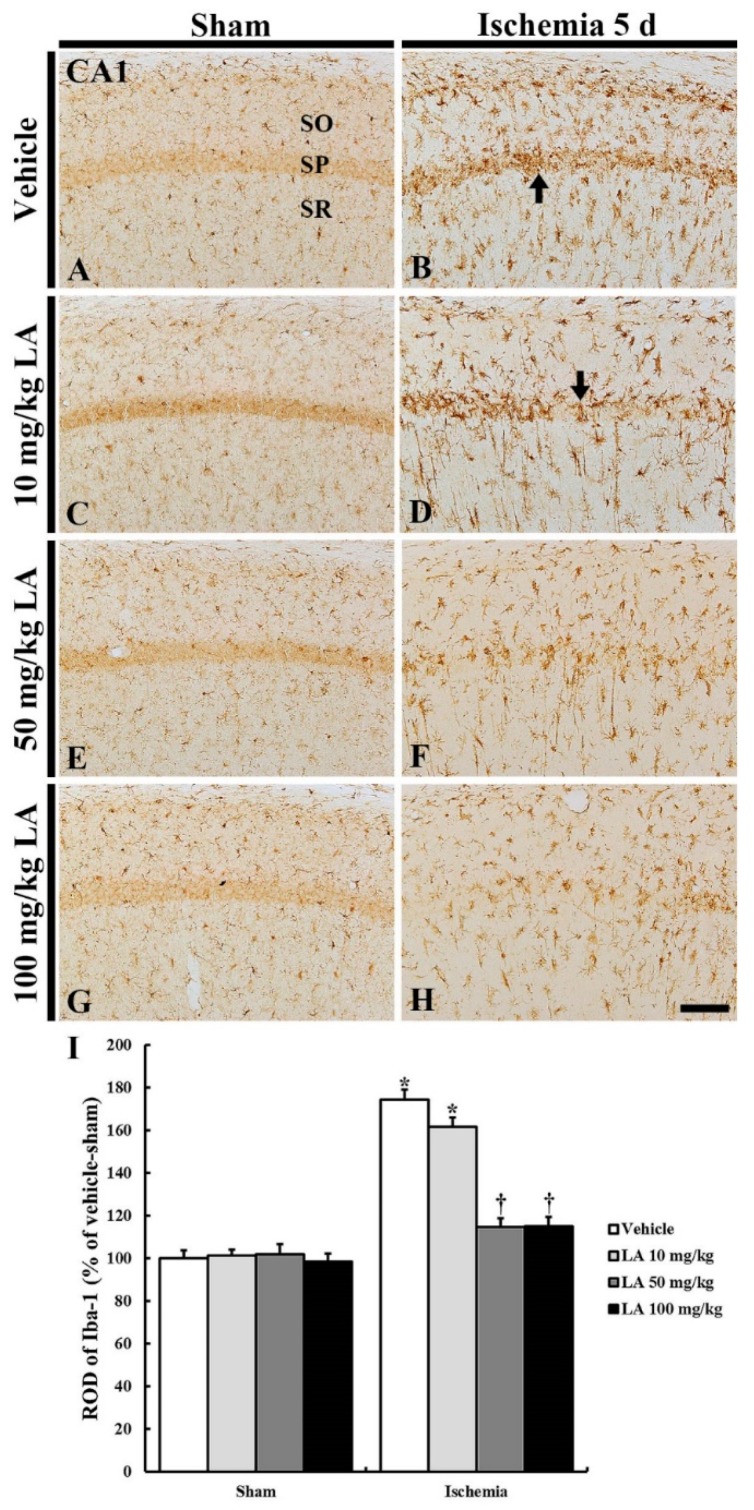
Figure 5.
Ionized calcium-binding adapter molecule 1 (Iba-1) immunohistochemistry in the CA1 field of the vehicle/sham (A), 10, 50 and 100 mg/kg LA/sham (C,E,G), vehicle/ischemia (B) and 10, 50 and 100 mg/kg LA/ischemia (D,F,H) groups at 5 days after sham or TFI operation. Iba-1 immunoreactive microglia are in a resting state in all the sham groups. In the vehicle/ischemia and 10 mg/kg LA/ischemia groups, Iba-1 immunoreactive microglia are hypertrophied, showing that many activated microglia gather in the SP (arrows). In the 50 mg/kg and 100 mg/kg LA/ischemia groups, activation of Iba-1 immunoreactive microglia is markedly attenuated, showing that they are evenly distributed in the CA1 field. Scale bar = 100 μm. (I) ROD (percentage) of Iba-1 immunoreactive structures in the CA1 field at 5 days after TFI (n = 7 in each group, * p < 0.05 versus vehicle/sham group, † p < 0.05 versus vehicle/ischemia group). The bars indicate the means ± SEM.

