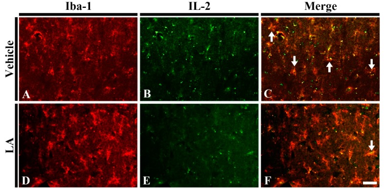
Figure 6.
Double immunofluorescence staining for Iba-1 (red), interleukin 2 (IL-2) (green) and merged images in the hippocampal CA1 field of the vehicle/ischemia (A–C) and 50 mg/kg LA/ischemia (D–F) groups at 5 days after TFI. Many IL-2 immunoreactive microglia (arrows) are shown in the vehicle/ischemia group. However, in the 50 mg/kg LA/ischemia group, a few IL-2 immunoreactive microglia are detected. Scale bar = 40 μm (n = 7 in each group).

