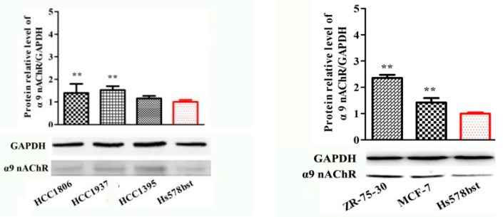
Figure 4.
Quantitative analysis of α9-containing nAChRs by Western blot in various cancer cell lines and breast epithelial cell lines. GAPDH was the control in all experiments. The proteins were detected with the corresponding antibody and an anti-mouse IgG secondary antibody conjugated to horseradish peroxidase (HRP). Each bar is the mean ± SD, n = 3. * p < 0.05, ** p < 0.01, breast cancer cell lines vs. breast epithelial cell lines HS578BST (normal cell control).


