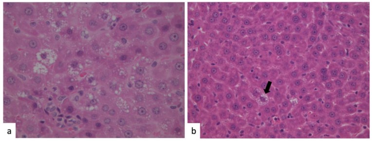
Figure 2.
Liver histology. Hematoxylin–eosin (H&E) staining of liver tissues obtained from rats fed with HFD (a) and HFD with algal phytocomplex (b). A diffuse microvescicular steatosis could be observed in rats fed with HFD (a), whereas very rare cells with microvescicular steatosis could be evidenced in the rats treated with the algal extract ((b), black arrow).

