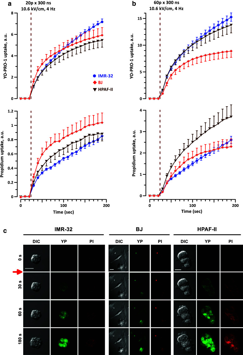
Fig. 6.
nsPEF permeabilizes cell membrane similarly in nsPEF-sensitive and nsPEF-resistant cell lines. Electroporative entry of marker dyes YO-PRO-1 (YP) and propidium iodide (PI) was monitored by time lapse fluorescence imaging in cell lines with high, intermediate, and low sensitivity to killing by nsPEF (IMR-32, BJ, and HPAF-II, respectively). A train of either 20 (a) or 60 (b) 300-ns pulses (10.6 kV/cm, 4 Hz) was delivered starting at 24 s into the experiment (vertical dashed line). Mean ± SEM for 14–34 cells in each group. The observed differences in dye uptake are not indicative of a stronger membrane permeabilization in more sensitive cell lines. Control cells showed no appreciable dye uptake within 200 s of the experiment (data not shown). c Representative images of morphological changes and dye uptake for exposures quantified in a. Red arrow indicates when the pulses were delivered. Scale bars are 20 µm. Note the enhanced blebbing in HPAF-II cells

