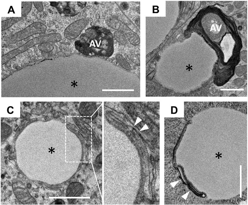
Figure 3. A subpopulation of CLDs has interconnections with AVs or is associated with membrane structures as observed by TEM.
(A and B) CLDs have physical contact with AVs. (C and D) Membrane structures are associated with the surface of CLDs. The images were taken from the enterocytes of Dgatl−/− mice 2 h after a dietary fat challenge. * = CLD; white arrows indicate membrane structures. Scale bar = 1 μm.

