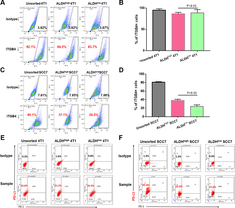
Figure 1.
Expression of ITGB4 and PD-L1 on 4T1 and SCC7 unsorted cells, ALDHhigh cells and ALDHlow cells. A: 4T1 cells highly expressed ITGB4 in unsorted, ALDHhigh and ALDHlow cell populations. B: Statistical data showed no difference in the expression of ITGB4 in ALDHhigh and ALDHlow 4T1 sub-populations (p>0.05). C: SCC7 cells also expressed high level of ITGB4 on unsorted cells, and its expression was significantly higher on ALDHhigh CSCs than on ALDHlow non-CSCs. D: Statistically, ITGB4 cell-surface abundance on ALDHhigh SCC7 cells was approximately 1.5-fold higher than that on ALDHlow SCC7 cells (p<0.05). E-F: PD-L1 expression on the 4T1 (E) and SCC7 (F) cells. Flow cytometry performed at least twice, and one presesentitive datum is shown for each experiment.

