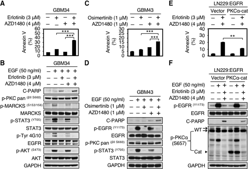
Figure 5.
EGFR and JAK2 inhibitors in combination induce apoptosis in part through inhibition of PKCα. A, GBM34 cells were treated with erlotinib, AZD1480, or erlotinib plus AZD1480 at indicated doses for 72 hours. Apoptosis was analyzed by flow cytometry for annexin V. Data shown represent mean ± SD (percentage of apoptotic cells relative to DMSO-treated control) of triplicate measurements (Student’s t test, p = 0.0002, DMSO versus erlotinib plus AZD1480; p = 0.0004, erlotinib versus erlotinib plus AZD1480; p = 0.0005, AZD1480 versus erlotinib plus AZD1480). B, An aliquot of each lysate was analyzed by western blot with antibodies indicated. EGF (50 ng/ml) was added 15 minutes before harvest. C, GBM43 cells were treated with osimertinib, AZD1480, or osimertinib plus AZD1480 at indicated doses for 48 hours. Apoptosis was analyzed by flow cytometry for annexin V. Data shown represent mean ± SD of triplicate measurements (Student’s t test, p = 0.0083, DMSO versus osimertinib plus AZD1480; p = 0.0005, osimertinib versus osimertinib plus AZD1480; p = 0.0001, AZD1480 versus osimertinib plus AZD1480). D, An aliquot of each lysate was analyzed by western blot with antibodies indicated. EGF (50 ng/ml) was added 15 minutes before harvest. E, LN229:EGFR cells were transduced with empty vector, or a dominant-active allele of PKCα (PKCα-Cat). Cells were treated with 3 μM erlotinib and 4 μM AZD1480 for 48 hours, Apoptosis was analyzed by flow cytometry for annexin V. Data shown represent mean ± SD (percentage of apoptotic cells relative to DMSO-treated control) of triplicate measurements {Student’s t test, p = 0.0007, DMSO (vector) versus erlotinib plus AZD1480 (vector); p = 0.0005, DMSO (PKCα-Cat) versus erlotinib plus AZD1480 (vector); p = 0.0018, erlotinib plus AZD1480 (PKCα-Cat) versus erlotinib plus AZD1480 (vector)}. F, An aliquot of each lysate was analyzed by western blot with antibodies indicated. EGF (50 ng/ml) was added 15 minutes before harvest.

