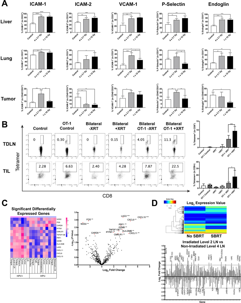
Figure 2:
Radiation modulates endothelial cell adhesion molecules and chemokines to enhance T-cell migration and preferential homing to irradiated sites. A) Percentage of cell adhesion molecules in CD45–CD31+ endothelial cells. (upper and middle) Naïve C57BL/6 mice received chest/abdominal irradiation (4 × 3.7 Gy or 1 × 12 Gy), and then the liver and lung were harvested 48 hours after the last irradiation. (lower) AT-84-E7 tumors in the flank were irradiated (4 × 3.7 Gy or 1 × 10 Gy) on Day 18, and tumors were harvested 48 hours after the last irradiation. Experiments repeated 3 times with similar results. B) Antigen-specific CD8+ T-cells in TDLN (upper) or TIL (lower) were detected by H-2Kb/SIINFEKL tetramer. Mice were inoculated with 1.5 × 10⁵ B16-OVA into unilateral or bilateral flank, and tumors on one side were irradiated (12 Gy) on Day 14. After 48 hours, 4 × 10⁶ T-cells from OT-1 mice were injected intravenously. Antigen-specific T-cells from both the irradiated tumor and contralateral tumor were analyzed 3 days after adoptive transfer. Data are shown as mean ± S.E.M. C) Differently expressed genes in 17 patient-derived HNSCC tissue samples comparing HPV-positive patients to HPV-negative patients analyzed using NanoString technology. D) Expression changes in mRNA detected by NanoString comparing an irradiated level 2 LN to a non-irradiated level 4 LN from the same patient. (Top) Heat map of the log2 value comparison between irradiated LN and non-irradiated LN mRNA. ≤5 (blue) shows low expression and ≥15 (red) showing high expression. (Bottom) Log2 fold changes of mRNA expression comparing an irradiated level 2 LN to a non-irradiated level 4 LN. mRNA Expression fold changes of ≥1.5 or ≤−1.5 are shown.

