Figure 5:
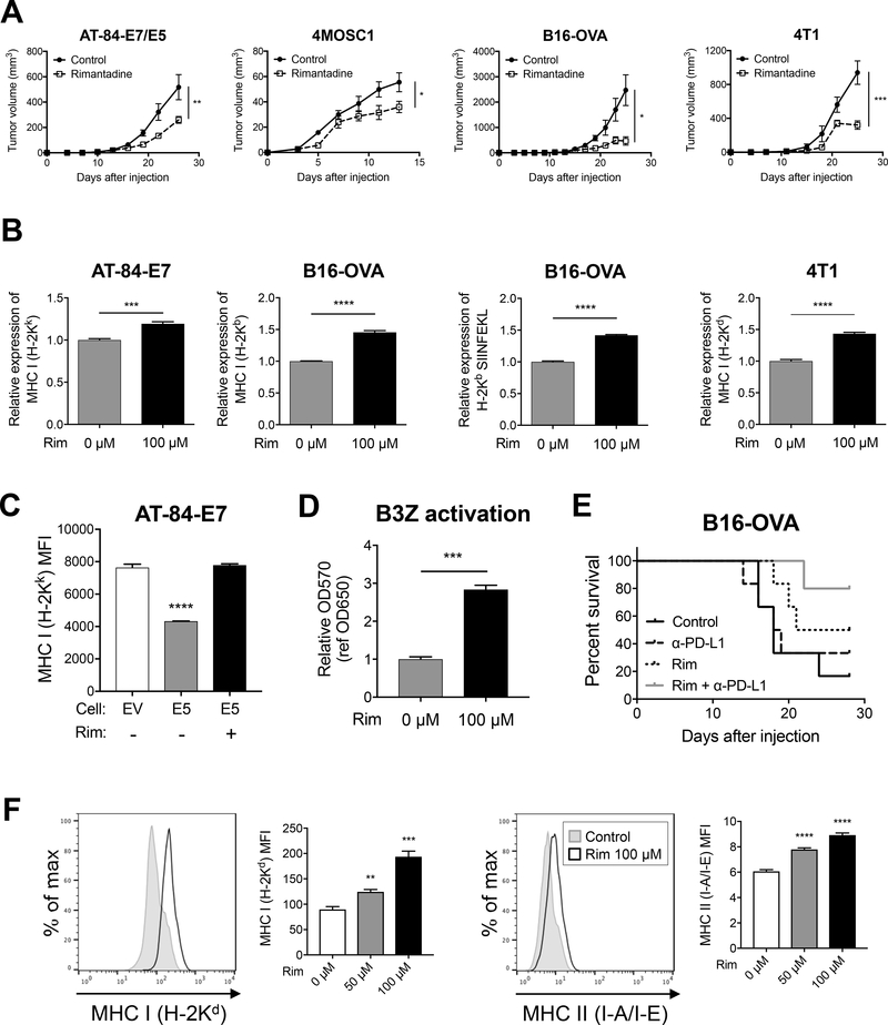
Rimantadine has novel anti-tumor activity and enhances MHC expression on tumor cells and antigen presenting cells. A) Tumor growth of AT-84-E7/E5, B16-OVA, and 4T1 (flank), and 4MOSC1 (tongue); n = 6 in each group. B, C) MHC I expression and antigen-presentation (H-2Kb/SIINFEKL) were analyzed by flow cytometry after rimantadine treatment (100 μM, 48 hours). D) Antigen-specific T-cell activation was analyzed by using B3Z after rimantadine treatment (48 hours). E) Survival curve of B16-OVA-bearing mice treated with rimantadine and/or anti-PD-L1 antibody; n = 6 in each group. F) Cell surface expression of MHC I (H-2Kd) and MHC II (I-A/I-E) on rimantadine-treated RAW264.7 48 hours after treatment. Data are shown as mean ± S.E.M.

